


Research Interests
The main focus of my research is problems in image analysis and pattern classification/recognition. In particular, I have experience with problems arising in clinical diagnostic imaging, seismic imaging, and computer vision. My research draws ideas from the following areas of applied and computational mathematics:
- Applied Harmonic Analysis, Wavelet Analysis, and Sparse Representations.
- Frame theory.
- Differential Geometry and Calculus of Variations.
- Probability and Statistics - Markov Random Fields.
I received my PhD in applied mathematics in August 2009, under the guidance of Prof. Manos Papadakis at the University of Houston, Department of Mathematics. My thesis deals with the development of isotropic wavelet frames for directionally unbiased processing of medical and seismic data.
Later as a post-doctoral research fellow at the University of Houston, I worked with Prof. Robert Azencott and Prof. Manos Papadakis on problems in biomedical imaging. We worked on rigid motion invariant classification of 3-D textures, and geometric modeling of the human mitral valve.
Currently, I am an assistant research scientist at Johns Hopkins University working with Prof. Michael Miller and Prof. Laurent Younes. I have developed an object oriented code for a diffeomorphic registration algorithm called Large Deformation Diffeomorphic Metric Mapping (LDDMM) , which lies at the core of the exciting field of Computational Anatomy.
Project Summaries
LDDMM and Computational Anatomy.
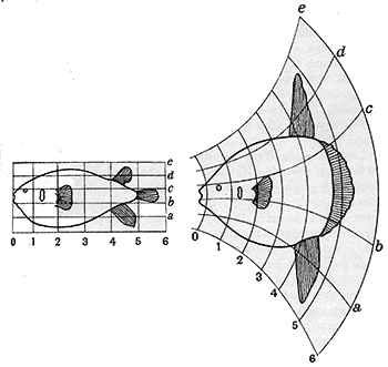
Computational Anatomy characterizes the anatomical shapes via the action of deformations on templates or exemplars. The statistical variabilities are then studied using probability models on the set of deformations. A Bayesian classification and testing approach can be used for the detection of anomalies based on these deformations.The idea of studying anatomy via transformations dates back to the work of D'Arcy Thompson's "On Growth and Form" published in 1917 (see figures below). A rigorous mathematical formulation of Thompson's idea was developed by Grenander and Miller following Grenander's global pattern theory.
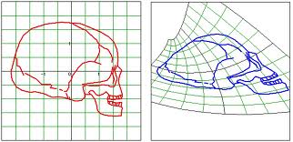
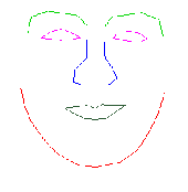
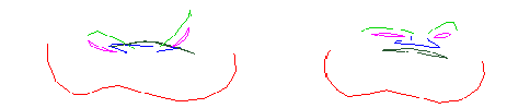
In the LDDMM framework, the deformations are diffeomorphisms which are smooth functions with smooth inverses. The following figure shows the advantage of using diffeomorphisms when dealing with large deformations. While a deformation of the top "face" using a diffeomorphism (bottom right) preserves the topology, a non-diffeomorphic transformation (bottom left) completely distorts it.
I have developed an object model for LDDMM and implemented it in C++, which facilitates fast implementation and experimentation for various modalities describing the anatomy, including curves, surfaces, and landmarks.
In particular, we have implemented a scheme to match a surface to cross sectional curves. This is used to study changes in shape of the left ventricle of the human heart due to cardiomyopathy. To obtain a higher temporal resolution, the MRI scans used in this study have a lower out of plain resolution. Hence, we only obtain sparse cross sections of the target surface. The figure below shows the template mesh (left) and the target curves (in red), and the mapped template (right).
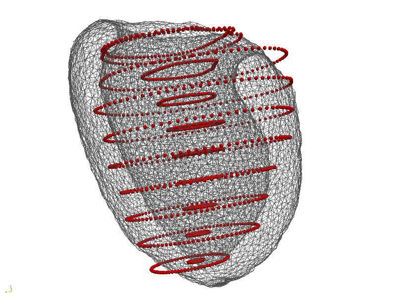
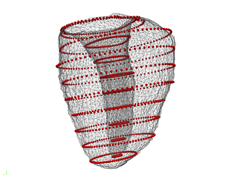
Using the LDDMM approach, left ventricle shape is parameterized by the "momentum" associated with the optimal deformation of the template. This enables statistical comparison of shape between diseased and healthy subjects using only the sparse cross-sections.
Isotropic Wavelet Transforms.
The ability to process a signal at multiple levels of resolution is highly desirable in signal and image processing as it allows us to adaptively process only the relevant details for a given problem. Wavelets, particularly those arising from Multiresoltion Analyses (MRAs), provide a computationally efficient multiscale signal representation. To date, the most popular examples of higher dimensional MRAs have been obtained from one dimensional MRAs via a tensor product construction because of the easy implementation of the associated wavelet transforms, which essentially involves the application of the 1D transforms along each dimension of the higher dimensional data. However, this very advantage is also one of the major drawbacks of this approach since it gives rise to what is known as directional bias. Isotropic Multiresolution Analyses (IMRA), constructed using radial filters, remedy this problem.

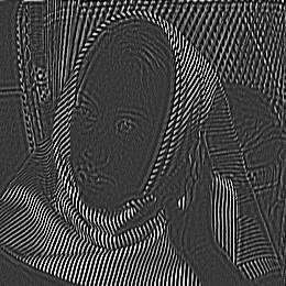
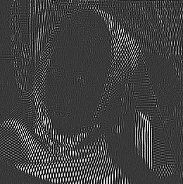
The image on the left is a standard image used in the image processing literature known as "Barbara". The center image is the high pass component using the isotropic wavelet filters, with texture at all orientations preserved, while it is highly distorted in the combined high pass channels of the tensor product Daubechies wavelet transform shown in the right image.
Segmentation of biomedical image data for automatic detection and recognition of anomalies.
In this project we used the isotropic wavelets arising from IMRA for the detection of atherosclerotic plaque in high resolution micro-CT scans of coronary arteries. The prediction filters are trained on the wavelet coefficients of normal tissue regions selected by experts. The classification step then classifies the regions whose statistics do not match those of the normal tissue. This is done by choosing thresholds on the response of normal tissue to the prediction filters.
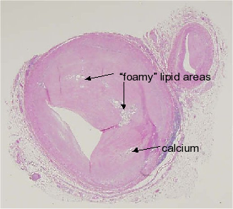
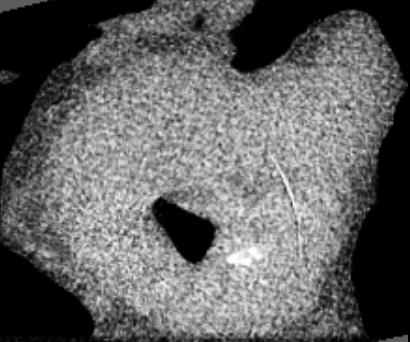
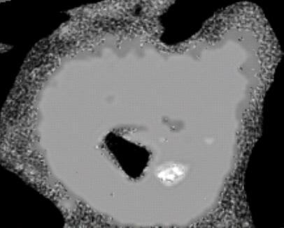
The left image is the result of histology, while the middle image shows unprocessed CT-data for the same slice. The image segmented by our algorithm shows the lipid region in light gray consistent with histology.
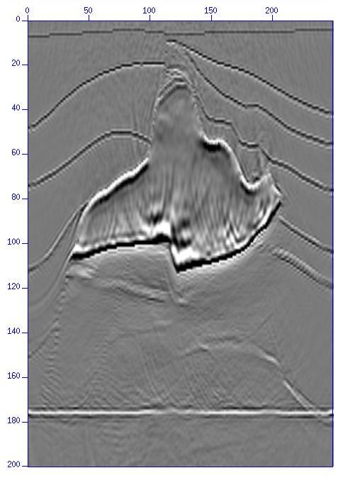
Numerical solution of the Acoustic Wave Equation in the context of Seismic Migration.
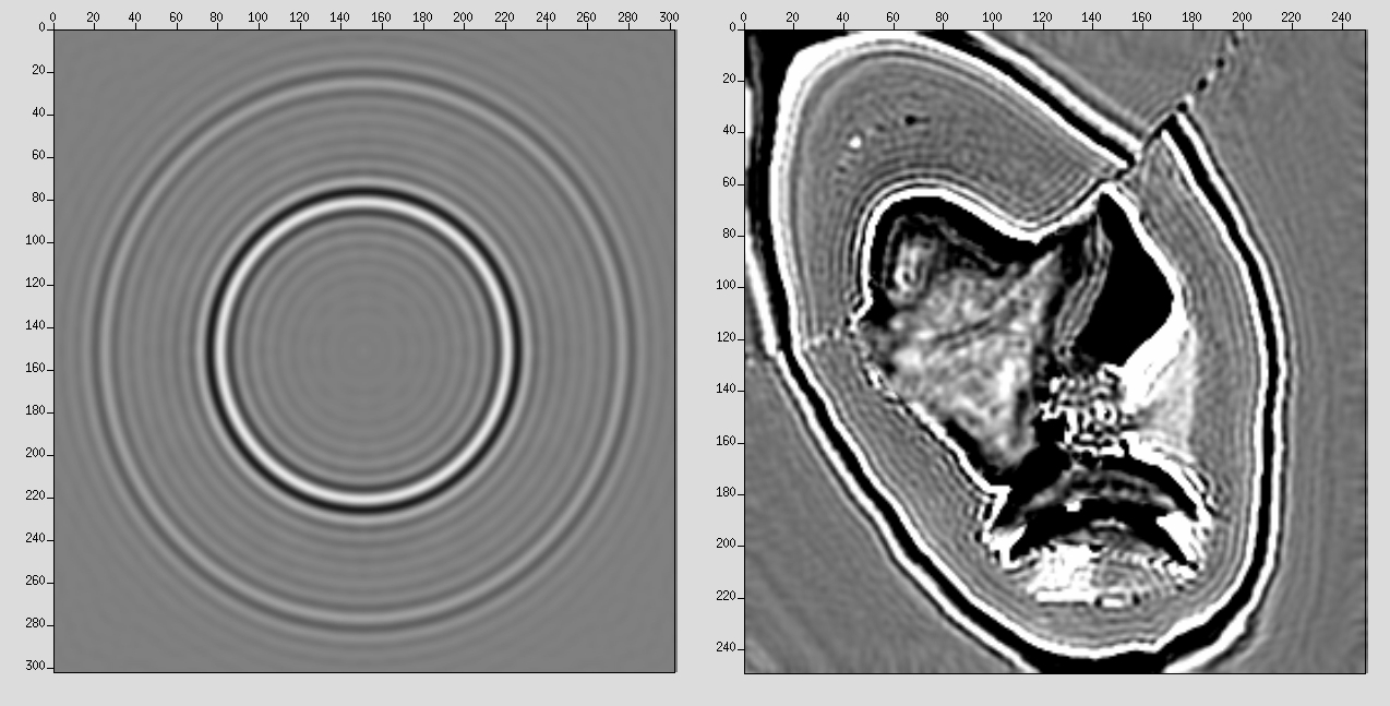
Migration is a seismic imaging technique used by the oil industry to image the interior of the Earth for the purpose of oil prospecting. A popular migration scheme, known as wave equation migration, uses the so-called Phase-shift propagator operator. The downward propagation of waves recorded on the surface by the recursive application of this operator constitutes an explicit scheme. Our goal was an efficient discretization of this operator using the isotropic wavelet transforms arising from IMRA, and using the multiscale structure to reduce the computational time of a standard explicit scheme. This project was funded by Total E & P, USA. The left and middle images below show horizontal cross-sections of the impulse response and the salt model. We see that the IMRA filters image well in all directions. The right image is a vertical cross section of the salt model, where we see superior image quality beneath the salt, which is difficult to achieve due to the high density of the salt dome.
Rigid Motion Invariant Classification of 3D textures.
In this project, we are interested in rotationally invariant classification of 3D textures because in applications such as medical imaging, a tissue type must be classified regardless of the position or orientation of the subject. Various groups have considered different approaches to obtain a rotationally invariant classification scheme, mostly in 2D. The main shortcoming of these models is that they either impose an isotropic structure on possibly non-isotropic textures, or can not be extented to 3D in a computationally efficient manner.
In this work we consider two different non-isotropic textures that might not be distinguishable if isotropy of the texture signatures is imposed. Hence, instead of using an isotropic texture model, we define a distance between two possibly non-isotropic texture signatures, which identifies a texture with all its rotations. We use the parameters of a Gaussian Markov Random Field fitted to the texture as feature vector, and the Kullback-Liebler divergence as a measure of "distance" between features. The rotation of discrete textures is implemented using an IMRA described above.
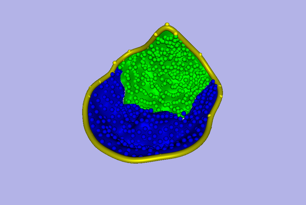
Geometric modeling of the human mitral valve from 3D-echocardiographic movies.
During my term as a Post-doc at UH, I worked with a team of cardiologists and mathematicians on the problem of matching deformable shapes in 3D-echocardiographic image sequences of the human heart. Our team developed tools for interactive tagging and modeling of the human mitral valve. The valve consists of two leaflets that guard the opening, and a fibrous ring surrounding the opening known as the mitral valve annulus. See figure below for a schematic of a mitral valve, and one of our models using NURBS.
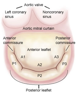
The medical community uses various geometric measurements such as the annular area, diameter and height of the mitral annulus to compare the geometry of a normal valve with a diseased valve. We complete these classical descriptors with the intrinsic concepts of curvature and torsion of a curve, to describe the geometry of the mitral annulus, and thus, give a more accurate description of the geometry independent of the coordinate system. These new measurements can lead to further refinements in mitral valve surgery to restore normal mitral valve geometry and function.
Further, the project aims at quantifying the static and dynamic stress on the mitral valve leaflets amongst patients with organic mitral regurgitation, to help surgeons plan better surgery, and prevent relapse of the disease.